How the Heart Beats
Clear, easy-to-follow explanations of how the heart functions and why that understanding matters.
UPDATED: 20 January 2026
Every moment of your life, your heart is quietly at work.
While you read this sentence, it is squeezing, relaxing, and refilling — pushing blood through a vast network of blood vessels that reaches every corner of your body. It does this without rest, without conscious effort, and usually without drawing much attention to itself.
Yet as we get older, small changes in how the heart works can have a big impact on our health. Understanding how your heart beats — not in medical jargon, but in plain language — can help you make sense of things like blood pressure, heart rhythm, and why doctors care so much about both.
This page will guide you through the heartbeat step by step. You don’t need any medical knowledge — just curiosity about how your body works and how to take better care of it.
Before we look at the individual parts of the heart, it helps to see the whole system in action.
In this short video, you’ll see a human heart beating inside the chest. That movement you can see is not random — it is the result of electrical signals, muscle contractions, and pressure changes working together in a precise and repeating pattern.
As you watch, notice how steady and rhythmic the movement is. Each beat follows the same sequence, over and over again, thousands of times a day.
In the sections that follow, we’ll break down exactly what is happening during each of those beats — and why it matters.
Video showing the heart beating inside the chest with a steady and rhythmic movement.
Heart anatomy: A guided tour inside your heart
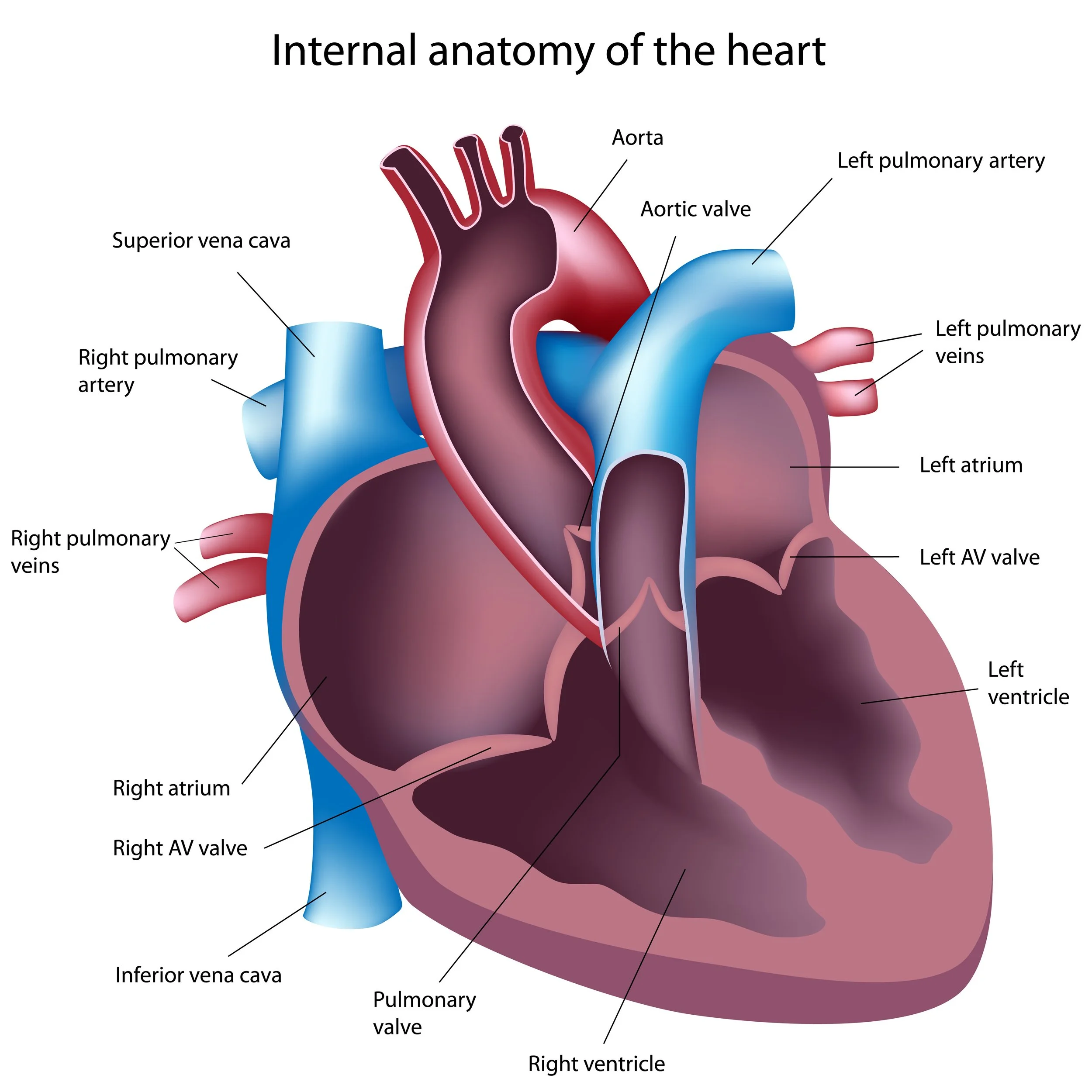
To understand how your heart beats, it helps to know how it is built.
Rather than thinking of the heart as a single solid muscle, imagine it as a small house with four rooms, one-way doors, and its own fuel supply. Each part has a specific role, and all of them must work together smoothly for every heartbeat to happen.
The four chambers – The heart’s rooms
A normal heart is divided into four chambers, each of which has a specific role.
The two upper chambers are called the atria. Their main job is to receive blood:
The right atrium collects blood returning from the body.
The left atrium collects blood returning from the lungs, freshly topped up with oxygen.
Below them are the two lower chambers, called the ventricles. These are the heart’s main pumping chambers:
The right ventricle pumps blood to the lungs.
The left ventricle pumps blood out to the rest of the body.
Although all four chambers are important, the ventricles do the hardest physical work — especially the left ventricle.
Interesting fact:
The left ventricle generates enough pressure to push blood all the way from your heart to your toes — and back again.
One-way valves – The heart’s doors
Between each chamber are valves, made of strong but flexible tissue.
These valves act like one-way doors. They open to allow blood to move forward and close to stop it flowing backward. They respond automatically to pressure changes inside the heart, opening and closing at exactly the right moments during each heartbeat.
When the valves are working well, blood flows smoothly and efficiently through the heart.
The left ventricle – The main pump
The left ventricle is usually the largest and most muscular chamber of the heart.
Its job is demanding: it must pump oxygen-rich blood out of the heart and into the arteries that supply your brain, organs, muscles, and other tissues. To do this, it has to generate enough force to push blood around the entire body.
Because of this workload, the muscle wall of the left ventricle is thicker than the walls of the other chambers.
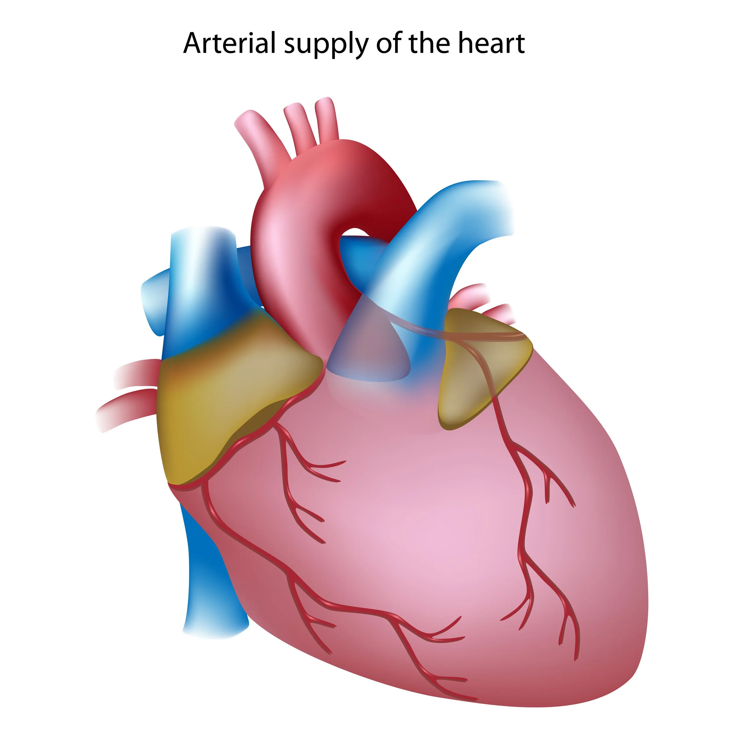
The heart’s own blood supply
Like any muscle, the heart needs its own supply of oxygen and nutrients to keep working.
This is delivered by the coronary arteries, which branch off from the body’s main artery (aorta) and wrap around the outside of the heart. They supply blood directly to the heart muscle itself.
If these arteries become narrowed or blocked, the heart muscle may not get the oxygen it needs — which can affect how well the heart works.
So far, we’ve looked at the different parts of the heart — its chambers, valves, and blood supply. But structure alone doesn’t make the heart beat.
For the heart to work as a pump, its muscle needs clear instructions about when to squeeze and when to relax. That timing comes from a built-in electrical system that quietly coordinates every heartbeat.
Electrical stimulation: What tells the heart when to beat
The heart’s natural pacemaker
Each electrical signal begins in a small area high in the heart, often described as the heart’s natural pacemaker.
From here, the signal spreads across the two upper chambers, causing them to contract and gently push blood down into the lower chambers.
This first step prepares the heart for the main pumping action that follows.
A brief pause, then a powerful squeeze
After the upper chambers have contracted, the electrical signal reaches a control point between the upper and lower chambers.
Here, there is a very short pause. This pause is important — it allows the lower chambers time to fill fully with blood before they contract.
Once past this point, the electrical signal travels rapidly through the walls of the lower chambers, triggering a strong, coordinated squeeze that pushes blood out of the heart and into the arteries.
In the video beside this text, you can follow this pathway step by step as the signal moves through the heart.
The heart does not beat by chance.
Every heartbeat is triggered by a small electrical signal that tells the heart muscle when to contract and when to relax. Without this signal, the heart muscle would not squeeze in a coordinated way.
This built-in electrical system works automatically, from birth to old age, keeping your heartbeat steady whether you are asleep, walking, or exercising.
Video showing each electrical stimulation starts high in the heart, spreading across the atria before pausing very briefly at a control point between the upper and lower chambers. Once past this point, the electrical signal travels rapidly through the walls of the ventricles.
Ready for the next beat
After each electrical signal, the heart briefly recharges.
Then the next signal begins, following the same pathway again. This repeating electrical pattern is what gives the heart its steady rhythm, beat after beat.
Most of the time, you’re completely unaware this system is working — which is exactly how it should be.
Electrical signals on their own don’t move blood. Their job is to trigger something far more powerful.
Once the timing is set by the electrical system, the heart muscle responds by squeezing and relaxing in a precise sequence. It’s this muscular action that turns electrical signals into movement — and movement into circulation.
Muscle contraction: How the heart pushes blood around your body
Knowing how the heart is built is only part of the story. What really matters is what happens each time it beats.
The heart is a powerful muscle. With every heartbeat, its muscular walls tighten, squeeze, and then relax again. This repeating cycle of contraction and relaxation is what keeps blood moving continuously through your body.
When the heart muscle contracts, the chambers become smaller and blood is forced forward. When the muscle relaxes, the chambers open up again and refill with blood, ready for the next beat.
This entire sequence happens in a steady rhythm, beat after beat, day and night.
From squeeze to flow
During each heartbeat, the ventricles — the lower chambers of the heart — do most of the pumping work.
As the ventricles contract, blood is pushed out of the heart and into the arteries. These arteries then carry the blood away, delivering oxygen and nutrients to your brain, organs, muscles, and tissues.
This movement of blood is not gentle. It requires force — and that force creates pressure inside the arteries.
Feeling your pulse
You can feel the effect of each heartbeat without any equipment.
When the heart pumps, it sends a pressure wave along the arteries. This wave is what you feel as your pulse.
You can check your pulse in several places where an artery runs close to the skin, such as at the wrist or on the side of the neck, as shown in the images.
If you place two fingers lightly on one of these spots, you’re not feeling the heart itself — you’re feeling the surge of blood caused by each contraction of the heart.
Did you know:
With each beat, the heart pumps roughly 70 millilitres of blood, which equates to 4 to 8 litres every minute, depending on heart rate — over the course of a day, that adds up to thousands of litres.
Video showing blood flow through the heart. Blood fills the atria before flowing into the ventricles. The ventricles soon contract, pumping blood out of the heart.


A moment to try it yourself
Try this now:
Sit quietly for a moment and gently feel your pulse. Notice how regular it is, and how each beat feels much the same as the one before it.
That steady rhythm is the result of your heart muscle contracting and relaxing in a carefully coordinated way.
Each squeeze of the heart doesn’t just move blood forward — it also raises the pressure inside the arteries.
That pressure rises and falls with every heartbeat, forming what we call blood pressure.
Understanding where this pressure comes from is the key to understanding why it matters so much to long-term health.
Blood pressure: Why each heartbeat creates pressure
Blood pressure is not something separate from the heartbeat — it is a direct result of it.
Each time the heart’s main pumping chamber contracts, blood is pushed into the arteries. Because these arteries are already full of blood, this push creates a rise in pressure inside them. When the heart relaxes again, that pressure falls.
This repeating rise and fall in pressure happens with every heartbeat, all day and night.
Two numbers, one cycle
Blood pressure is usually described using two numbers, which reflect two different phases of the heartbeat.
Systolic blood pressure is the higher number. It represents the pressure in the arteries at the moment the heart contracts and pushes blood out.
Diastolic blood pressure is the lower number. It represents the pressure in the arteries while the heart is resting and refilling between beats.
Together, these numbers give a picture of how hard the heart is working to move blood around the body.
Worth knowing:
Many people with high blood pressure feel completely normal — which is why regular measurement is so important.
Why blood pressure matters
Blood pressure needs to be high enough to keep blood flowing to vital organs like the brain and kidneys. But if it is consistently too high, it places extra strain on the heart and blood vessels.
Over time, this added strain can affect how well the heart works and increase the risk of health problems — often without causing any obvious symptoms at first. Persistently raised blood pressure, also known as high blood pressure or hypertension, is common as we get older.
This is why blood pressure is sometimes described as a “silent” issue: you may feel perfectly well, even when it is higher than it should be.
A link you can feel
The pressure created by each heartbeat is the same pressure wave you feel when you check your pulse. Although the pulse tells you how fast your heart is beating, blood pressure tells you how much force is being generated with each beat.
Both measurements come from the same heartbeat, but they provide different information.
Key Takeaways: How your heartbeat, pressure, and health are connected
The heart beats in a steady rhythm, guided by electrical signals.
Each heartbeat causes the heart muscle to squeeze and relax.
These squeezes create pressure inside the arteries.
This rise and fall in pressure is what we call blood pressure.
Over time, changes in blood pressure can affect how hard the heart has to work.
Blood pressure gives us valuable clues about how hard the heart is working. When it remains raised over time, the heart and blood vessels are placed under constant strain.
The next page looks at what happens when blood pressure stays higher than it should — a condition known as hypertension — what it is, how it develops, and why regular monitoring plays such an important role in protecting long-term health.
This page is part of a series designed to help you better understand your heart and blood pressure.
Important Information:
This page is provided for general educational purposes only and does not constitute medical advice. Medical knowledge and guidance evolve over time, and information may change. Always seek advice from a qualified healthcare professional regarding personal health concerns or medical decisions.
For more details, please see our full Disclaimer.